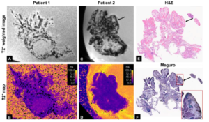
Figure 1. High resolution ex vivo MRI at 7T, histology, and iron correlation. A) Transverse view of fimbria at 9.42 ms echo time of a normal, resected fallopian tube from a patient with SDHB mutation. Transverse view demonstrates fine vessels as black stripes on the shortest echo time (160m resolution). B) Corresponding T2* map reconstructed from three echo acquisition. C-F) Patient with HGSCO treated with neoadjuvant chemotherapy prior to resection. C) Transverse view of fimbria at 9.42 ms echo time demonstrates more architectural distortions and more regions with pronounced T2*w contrast (dark regions, black arrow) at 240 m resolution. E) H&E stain. F) Meguro iron stains demonstrate areas of iron deposition (brown) distributing to areas of T2*w contrast in patient 2 (blackarrows). Inset (red box). Photo credit: Technion Spokesperson’s Office)
A research team led by Prof. Moti Freiman of the Technion Faculty of Biomedical Engineering and the May-Blum-Dahl Human MRI Research Center has received a prestigious Gray Foundation Team Science grant to develop an AI-powered MRI method for the early detection of ovarian cancer. The project is one of several selected as part of the Foundation’s latest $35 million investment in research aimed at preventing, intercepting, and detecting BRCA-related cancers at their earliest stages.
The project brings together two complementary research efforts: a Technion–University of Pennsylvania team led by Prof. Moti Freiman, Dr. Karthik Sundaram, and Prof. Dylan Tisdall, and a University of Chicago team led by Dr. Milica Medved and Dr. Kathryn Mills. Together, the teams will combine advanced MRI, quantitative imaging, and artificial intelligence to develop effective, affordable MRI-based screening strategies for ovarian cancer.
Ovarian cancer remains one of the deadliest gynecologic cancers because it is usually diagnosed only after it has spread beyond the ovaries. Women who carry inherited BRCA1 or BRCA2 gene mutations face a dramatically increased lifetime risk of developing the disease. As a result, many women with these mutations are advised to undergo preventive removal of their ovaries and fallopian tubes. This lifesaving but irreversible procedure has profound consequences, particularly for younger women.
The newly funded project aims to provide physicians and patients with a more precise way to assess cancer risk before such surgery becomes necessary.
The researchers have developed an innovative MRI protocol that detects elevated iron levels in the fallopian tubes, an early biological marker associated with the development of high-grade serous ovarian cancer, the most common and aggressive form of the disease. By identifying these changes at a precancerous stage, the technology could enable earlier diagnosis while improving clinical decision-making regarding preventive surgery.
The Technion’s contribution to the project centers on advanced MRI analysis powered by artificial intelligence. MRI scans of healthy volunteers will be conducted at the May-Blum-Dahl Technion Human MRI Research Center, where the project’s initial imaging data were also collected. The Technion team will apply AI algorithms developed in Prof. Freiman’s laboratory to identify imaging biomarkers that are difficult or impossible to detect using conventional image analysis. Previous studies by Prof. Freiman’s group have demonstrated the ability of these methods to uncover clinically meaningful biomarkers from MRI scans.
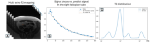
Figure 2. Multi-echo MRI signal decay and multicomponent T2 analysis pipeline. A) Representative multi-echo MRI acquisition with sequential echo images and a region-of-interest (ROI) in pelvic tissue of a healthy volunteer. B) Signal decay from the ROI showing measured data (points), multicomponent T2 fit (dashed), and conventional monoexponential (scalar) T2 fit (dotted). C) Corresponding T2 distribution demonstrating multiple relaxation components: short-T2 components likely reflect rapidly relaxing compartments (e.g., dense microstructure or susceptibility-related effects such as blood products), intermediate components reflect cellular and stromal tissue, and longer-T2 components reflect fluid-rich compartments. The vertical dashed line denotes the conventional monoexponential (scalar) T2 value. Photo credit: Technion Spokesperson’s Office)
At the University of Pennsylvania, researchers will perform MRI scans of surgically removed cancerous ovaries. These studies are currently conducted using a rare and costly 7 Tesla MRI system, which offers exceptionally high image resolution but is available at only a limited number of research centers worldwide. Together, the Penn and Technion teams will demonstrate that the same diagnostic information can be obtained using the much more common 3 Tesla MRI scanners found in hospitals around the world, paving the way for broad clinical adoption.
The University of Chicago team, led by Dr. Milica Medved and Dr. Kathryn Mills, will combine quantitative MRI and artificial intelligence to support the design of effective, affordable MRI screening strategies for ovarian cancer.
The Gray Foundation’s Team Science program supports multidisciplinary collaborations focused on preventing, intercepting, and detecting BRCA-associated cancers before they become life-threatening. The Foundation’s latest funding round awarded $35 million to leading research teams pursuing innovative approaches to breast, ovarian, pancreatic, and prostate cancers associated with BRCA mutations. If successful, the project could lead to a clinically accessible screening tool that enables earlier detection of ovarian cancer and helps women at high genetic risk make more informed decisions about preventive treatment.
The Gray Foundation is focused on accelerating research, improving treatment, and raising awareness for individuals who have inherited BRCA mutations. With these grants, the Gray Foundation has committed $235 million to support BRCA research.
For further reading, here