New materials developed by Technion researchers will assist in the early diagnosis of various diseases and reduce the need for radiation-intensive tests
The materials, which are expected to lead to significant improvements in the quality of Magnetic resonance imaging (MRI) scans and expand their usage, are presented in a newly published article by Prof. Aharon Blank and Dr. Itai Katz in Science Advances. The research, supported by the Technion Human Health Initiative (THHI) and the European Research Council (ERC), included contributions from Prof. Boaz Pokroy and Dr. Arad Lang from the Faculty of Materials Science and Engineering at the Technion, who worked on preparing some of the unique nature-inspired materials, and Dr. Benno Meier from the Karlsruhe Institute of Technology.

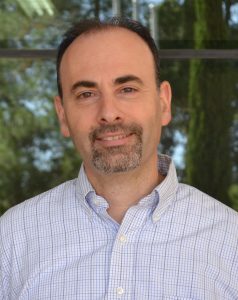
Prof. Aharon Blank (left) and Dr. Itai Katz
MRI is a non-invasive, radiation-free imaging technology used in diagnosing various clinical conditions. One of the limitations of conventional MRI devices is that they have trouble detecting metabolites, which have a very low concentration in tissues. Metabolites are small molecules involved in chemical process in the body, many of which serve as clinical markers indicating various health conditions, including malignant tumors, abnormal cell division, cell death, and cellular stress. This is the motivation for many research groups trying to find a solution that allows the identification of metabolites in non-invasive imaging scans.
In their article, the researchers present a new method enabling the identification of metabolites in MRI. The method, called MMV, is based on new composition of metabolites characterized by two significant advantages in this context: a dramatic enhancement (by about four orders of magnitude) of the magnetic resonance signal and the preservation of signal strength for a relatively long time compared to existing metabolites – about ten minutes versus one minute.

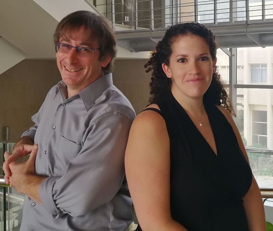
Dr. Arad Lang
Prof. Boaz Pokroy
The practical implication of the findings is that the new materials will allow tracking metabolites in various tissues over time. Furthermore, due to the new qualities these materials provide to MRI scans, such tests could, in certain cases, replace expensive and radiation-intensive tests like PET-CT.
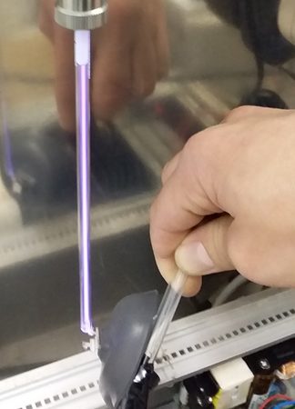
One of the stages in the preparation process of the unique materials. This stage includes high-voltage plasma disintegration passing through the material grains
According to Prof. Blank, “Our discovery is very exciting for us, as the new method will provide physicians with a broader time window to perform the scan, and we estimate it will expand the use of radiation-free MRI scans. These materials will improve the capabilities of medical and research teams in early disease diagnosis, tissue characterization, disease progression monitoring, surgery planning, optimal treatment selection, and informed decision-making.”
Click here for the paper in Science Adva